Ankle Anatomy 101: The Bones and Structures in Your Ankle
A plain-language tour of the bones that make up your ankle, why the anatomy explains so much about fractures, swelling, and healing, and how to think about what you're feeling when something goes wrong.

Most people don’t go looking for ankle anatomy out of curiosity. They go looking because something happened. The ankle rolled, or it got hit, or it swelled up overnight, or an X-ray report came back with words on it that nobody explained.
And anatomy, when it’s explained well, makes the whole thing less mysterious. If you know which bone is which, you understand why the outside of your ankle swells more than the inside. You understand why one fracture gets a boot and another gets surgery. You understand why the ankle can hurt in a spot two inches from where you thought the problem was.
So let’s walk through it. Not a textbook chapter. Just the parts that actually change how you think about your ankle.
What are the bones of the ankle called?
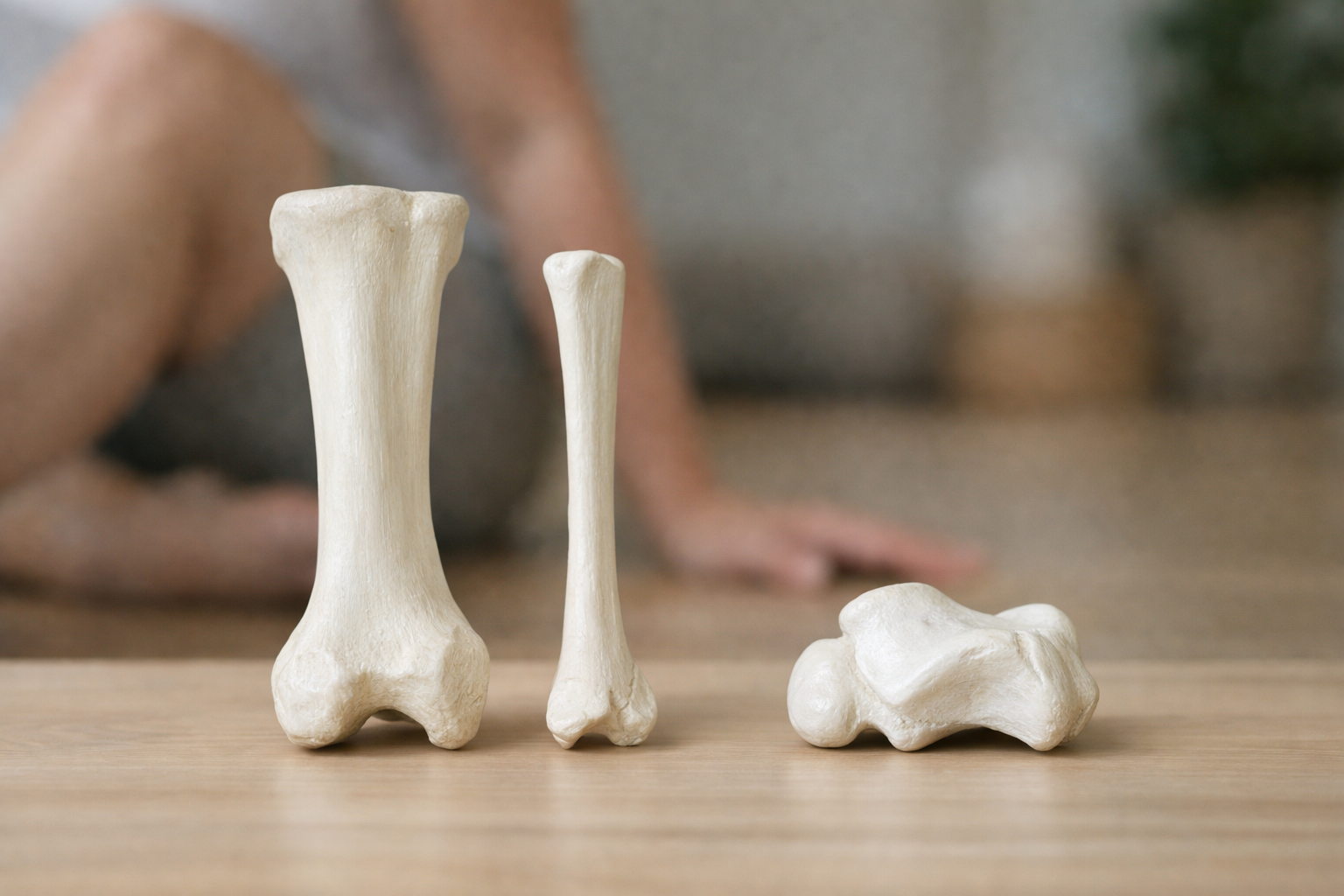
The ankle joint itself, the hinge that lets your foot point down and pull up, is built from three bones.
The tibia. Your shin bone. It’s the big weight-bearing bone of the lower leg, and it carries almost all of the load from your body down into your foot. The bottom end of the tibia forms the roof of the ankle joint, sometimes called the plafond, which is just an old word for ceiling. The tibia also forms the bump on the inside of your ankle, the medial malleolus.
The fibula. The thinner bone running alongside the tibia on the outside of your leg. It carries far less weight, maybe a small fraction of what goes through the tibia. Its job is more about stability and giving muscles and ligaments somewhere to attach. The bottom end of the fibula becomes the bump on the outside of your ankle, the lateral malleolus.
The talus. This is the one most people have never heard of, and it’s the interesting one. The talus sits underneath the tibia and fibula like a block wedged into a socket. It’s the bridge between your leg and your foot. Here’s what makes it unusual: no muscles attach to the talus. Not one. It’s held in position entirely by the shape of the bones around it and by ligaments. Everything that happens at your ankle passes through this bone.
Those three bones together form what’s often called the true ankle joint, or the tibiotalar joint. Below that sits a second joint, between the talus and the heel bone (the calcaneus), which handles the side-to-side tilting motion of your foot. When you walk on a slanted sidewalk and your foot adjusts to the angle, that’s mostly happening below the ankle joint proper, not in it.
That two-level design matters. The upper joint is a hinge: up and down. The lower joint is a swiveling, tilting joint. Together they let your foot handle ground that isn’t flat. When people say an ankle feels stiff after an injury, it’s often worth figuring out which of those two levels is actually stiff, because they respond to different things.
What are the 7 bones in the ankle area collectively called?
The seven bones in the back half of your foot are called the tarsal bones, or the tarsus. Some sources describe them loosely as “ankle bones” because they sit in the ankle region, which is where the confusion comes from. Two of them (the talus and the calcaneus) are directly involved in ankle motion. The rest are more accurately mid-foot bones.
Here they are:
- Talus – the bridge bone described above, sitting under the tibia and fibula.
- Calcaneus – your heel bone, the largest bone in the foot, and the anchor point for the Achilles tendon at the back and the plantar fascia underneath.
- Navicular – a boat-shaped bone on the inner side of the mid-foot; the posterior tibial tendon attaches here, which matters for people with arch problems.
- Cuboid – on the outer side of the mid-foot, roughly opposite the navicular.
- Three cuneiforms (medial, intermediate, lateral) – a row of wedge-shaped bones sitting between the navicular and the long bones of the foot. They form part of the arch structure.
Beyond the tarsals you have the five metatarsals (the long bones running toward your toes) and the phalanges (the toe bones). All in, the foot and ankle have 26 bones and more than 30 joints. That’s about a quarter of the bones in your entire body, packed into a structure the size of your shoe.
Which tells you something. The foot and ankle aren’t built to be rigid. They’re built to be adaptable, to change shape as you move across ground, to be soft and accommodating when your heel lands and stiff and springy when you push off. That’s a lot of moving parts, and it’s part of why foot and ankle problems can be hard to pin down without an in-person exam.
What is the bone that sticks out on the outside of your ankle?
That’s the lateral malleolus, and it’s the bottom end of your fibula.
Run your fingers over both sides of your ankle and you’ll notice something. The bump on the outside sits lower and further back than the bump on the inside (the medial malleolus, from the tibia). That asymmetry is not a design flaw. The outer bone extends further down to act like a wall, blocking the ankle from rolling outward.
It also explains a lot about injuries. Rolling inward, where the sole of the foot turns toward the other leg, is by far the more common direction, because there’s less bony blocking on that side. The ligaments on the outside of the ankle take the strain, and that’s why the outside is where most sprain pain and swelling shows up.
One more piece of anatomy worth knowing: the tibia and fibula aren’t just sitting next to each other. They’re bound together at the bottom by a set of ligaments called the syndesmosis. That connection keeps the two bones snug around the talus. When a twisting force pulls those two bones apart, you get what people call a high ankle sprain, and it tends to behave differently and take longer than a standard outside-ankle sprain. If your pain sits above the ankle joint rather than below the outer bump, that’s worth mentioning to whoever examines you.
What is the most common ankle bone to break?
The fibula, specifically the lateral malleolus at its lower end. It’s the most commonly fractured of the ankle bones by a wide margin.
That makes sense given the mechanics. The fibula is the thinner bone. Its lower tip sticks out furthest. It’s the structure resisting the most common direction of rolling. When a twist overwhelms the ligaments on the outside, sometimes the ligament tears, and sometimes it holds and the bone gives way instead.
Ankle fractures get grouped roughly by how many of the malleoli are involved:
- Unimalleolar – one bone broken, usually the fibula. Often stable, often treated without surgery.
- Bimalleolar – both the inner and outer bumps broken. This usually means the joint has lost its stability, and it more often needs surgical fixation.
- Trimalleolar – both malleoli plus the back edge of the tibia. More complex again.
The number of broken bones matters less than one question: is the joint still lined up and stable? A single clean crack in the fibula where everything stays in position is a very different situation from a single crack where the talus has shifted inside the joint. That’s what your surgeon or physician is really assessing when they look at your imaging. Not just “is there a break” but “will this joint stay where it belongs while it heals.”
What’s the worst bone to break in your foot?
Surgeons and therapists tend to worry most about fractures of the talus and the calcaneus.
The talus is difficult for a specific reason: its blood supply. Blood enters the talus in a somewhat roundabout way, and a significant fracture can interrupt that supply. When bone loses its blood supply, part of it can die off, which is called avascular necrosis. That can lead to collapse of the bone surface and arthritis in the joint. Talus fractures usually come from high-energy events, falls from height, car accidents, and they’re taken seriously.
The calcaneus is difficult for different reasons. It’s largely a shell of hard bone around a spongy interior, and when it breaks it often shatters rather than cracking cleanly. Because the heel bone sets the alignment for everything above it, a calcaneus fracture that heals in a poor position can change how you load your whole foot for years afterward.
A third one gets less attention but frustrates a lot of active people: the fifth metatarsal, particularly a break at a specific zone near its base sometimes called a Jones fracture. That area has a limited blood supply too, and those fractures are known for healing slowly or not fully knitting together.
None of this is meant to alarm you. Most ankle and foot fractures are not in this category. But it explains why two people can both say “I broke my ankle” and have completely different recoveries, and why comparing your timeline to a friend’s rarely tells you much.
Can you walk on a fractured ankle bone?
Yes, sometimes, and this is exactly why people delay getting checked.
Plenty of people walk into a clinic days after an injury, limping but walking, and leave with a fracture diagnosis. It happens most with small avulsion fractures (where a ligament pulls off a fleck of bone), with stable fibula fractures, and with stress fractures that build gradually rather than happening in one moment.
So being able to bear weight does not rule out a break. That’s the practical point. The ability to walk tells you something about pain tolerance and about how much of the load-bearing structure is still intact, but it isn’t a reliable test.
What should actually push you toward getting imaged:
- You couldn’t put weight on it at all immediately after the injury, and still can’t take four steps.
- There’s tenderness directly on the bone, on the back edge or tip of either ankle bump, rather than on the soft tissue nearby.
- There’s tenderness at the base of the fifth metatarsal (the bony bump halfway along the outer edge of your foot) or on the navicular.
- The ankle looks visibly out of shape, or the swelling and bruising are dramatic and immediate.
- Numbness, a cold or pale foot, or pain that feels out of proportion and keeps climbing.
Those bone-tenderness points aren’t arbitrary. Emergency clinicians use a structured set of criteria to decide who needs an X-ray after an ankle injury, and pressing on those specific spots plus checking weight-bearing is essentially it. If a couple of those apply to you, get it looked at. An X-ray is quick and settles the question.
How can I tell if my ankle is broken or fractured?
First, a small clarification that catches people off guard: broken and fractured mean the same thing. There’s no medical distinction. A hairline fracture is a break; a shattered bone is a break. The words describe severity, not two different categories of injury.
As for telling a fracture from a bad sprain, the honest answer is that you often can’t, not reliably, not from the outside. Severe sprains can swell more and hurt more than stable fractures. I’d rather tell you that plainly than give you a checklist that sounds more certain than it is.
That said, some patterns lean one direction:
Leans toward fracture:
- Pinpoint pain on the bone itself that you can cover with one fingertip
- A crack or snap sound at the moment of injury, especially with an immediate inability to weight-bear
- Swelling that appears within minutes and is severe
- Visible deformity, or the foot pointing at an odd angle relative to the leg
- Pain that doesn’t ease at all with rest and elevation over the first day or two
Leans toward sprain:
- Pain spread over a broader area of soft tissue, usually below and in front of the outer ankle bump
- Swelling that develops over a few hours rather than instantly
- You can bear weight, awkwardly, and it improves noticeably in the first few days
Even with those patterns, imaging is what settles it. And there’s a category that X-rays can miss entirely: stress fractures, which often don’t show up for two to three weeks until healing bone becomes visible. If you have an ankle or foot pain that came on gradually with increased activity, stays in one precise bony spot, and hurts more the longer you’re on it, a normal early X-ray doesn’t rule that out. That situation deserves a follow-up, not a shrug.
If you’ve had a sprain confirmed and you’re trying to figure out what comes next, we’ve written separately about how to treat a sprained ankle and the decisions around protecting versus moving it.
Can an ankle fracture heal without a cast?
Often, yes. Traditional plaster casts have become less automatic than they used to be.
What’s replaced them for many stable fractures is a removable walking boot, a walker boot, or a brace. For a stable fibula fracture where the joint is aligned and the talus hasn’t shifted, a boot with a defined weight-bearing plan is frequently enough. Some fractures need to be non-weight-bearing for a stretch, then progress. Some allow weight-bearing as tolerated from early on.
The deciding factor is stability, not comfort or convenience. Bone heals when the broken ends stay close together and reasonably still. A cast, a boot, or a plate and screws are three different ways of accomplishing the same thing. Which one you need depends on what the imaging shows and what your physician judges about the joint’s alignment.
What matters just as much, and gets talked about less, is what happens after the bone heals. Six weeks in a boot does a great deal to the calf muscle, the ankle’s motion, and your balance system. The bone knits and the imaging looks good, and then people are surprised that walking normally still feels hard. That gap between “healed” and “capable” is real, and closing it is a rebuilding process, not a waiting one. The bone heals on biology’s schedule. Everything around it, the muscle, the tendon stiffness, the sense of where your foot is in space, has to be rebuilt through gradually asking more of it.
What not to do with a fractured ankle
A few things genuinely work against you:
Don’t ignore weight-bearing instructions in either direction. If you’ve been told not to put weight through it, that restriction usually exists because the alignment is fragile. If you’ve been cleared to bear weight, avoiding it “just to be safe” isn’t safe. Bone responds to load. Prolonged unnecessary unloading weakens it.
Don’t leave the boot on 24/7 without asking about motion. Depending on the fracture, your physician may want the ankle completely immobilized, or may allow gentle ankle motion out of the boot at certain points. Ask. Weeks of total stillness cost you range of motion and calf muscle that take much longer to get back than they took to lose.
Don’t skip the follow-up X-ray because it feels better. Feeling better and being aligned are separate things. Fractures can shift in the early weeks without any dramatic pain signal.
Don’t assume you’re done when the boot comes off. This is the big one. Coming out of a boot, your calf will be smaller, your ankle stiffer, and your balance measurably worse than the other side. Walking normally on flat ground returns fairly quickly. Handling stairs, curbs, grass, a wet floor, or a sudden step off a sidewalk takes longer, because those things demand a lot more from an ankle than a level hallway does.
Don’t push through sharp, localized bone pain that’s getting worse week over week. General soreness and stiffness as you get moving again is expected and usually fine. A specific point on the bone that hurts more each week is worth a call.
That last distinction is worth sitting with, because it applies well beyond fractures. Discomfort during rehab is not automatically damage. Pain is information about how much your ankle was ready for that day, and it doesn’t always mean you undid something. For most people rebuilding after an injury, keeping discomfort at a manageable level (often somewhere around a 4 out of 10 or below during and after activity) and checking how it feels the next morning gives you a workable guide. Settled by the following day usually means the dose was reasonable. Still elevated 48 hours later usually means back off a notch, not stop.
Why any of this anatomy actually helps you
Knowing bone names doesn’t heal anything. But it changes how you interpret what you feel, and that matters more than people expect.
When you know the fibula carries relatively little weight, you understand why a stable fibula fracture can sometimes be walked on and a tibia fracture usually can’t. When you know the talus has no muscle attachments and depends on ligaments and bone shape for position, you understand why ankle stability work is largely about the structures around the joint. When you know there are two separate joint levels in the ankle, you can start noticing whether your stiffness shows up going up on your toes (upper joint) or walking across a slanted driveway (lower joint).
And when you know that the foot and ankle contain 26 bones and dozens of joints designed to adapt to uneven ground, it makes sense that recovery isn’t finished when flat-ground walking feels fine. The ankle has to become capable of the ground you actually live on.
Recovery gets built through that process. Load a little, watch what your ankle does with it, adjust, repeat. Some weeks are better than others, and a rough week doesn’t erase the previous six.
When to have someone look at it
See a physician promptly for an obvious deformity, a foot that’s numb, pale, or cold, an inability to bear any weight after an injury, or pain and swelling that keep worsening rather than settling over the first few days. Those need imaging and hands-on assessment, not a self-guided plan.
For ankle pain that’s lingered past a few weeks, or a fracture that’s healed but left you unsure how to get back to your actual life, an in-person evaluation is what sorts out which structure is involved and what your ankle is currently ready for. Not every ache in that region is what it first appears to be. Tendon problems, nerve irritation, and joint surface injuries can all masquerade as one another, and a description over the internet, however detailed, can’t replace someone putting hands on your ankle and watching you move.